最高のコレクション e coli gram stain shape 232883-E coli gram stain shape
What is the shape & arrangement of E coli?However, there are many differences exhibited between E Coli and Salmonella E Coli E coli is rather a common reference than a scientific name of the wellknown gramnegative bacteria that poisons the human food to dangerous consequences The scientific notation of this bacterium should be presented as the Escherichia coli or E coli, in the italicized letters E coli is a facultative anaerobic bacterium with a rodshaped bodyFor rodshaped or filamentous bacteria, length is 110 µm and diameter is 0251 0 µm E coli , a bacillus of about average size is 11 to 15 µm wide by to 60 µm long Spirochaetes occasionally reach 500 µm in length and the cyanobacterium Oscillatoria is about 7 µm in diameter
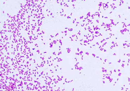
Gram Negative Bacteria Images Photos Of Escherichia Coli Salmonella Enterobacter
E coli gram stain shape
E coli gram stain shape-Gram stain results can assist in choosing an appropriate drug treatment shape, edge, elevation, color, odor, and texture is part of ___ A microbiologist inoculates Staphylococcus epidermidis and Escherichia coli into a culture medium Following incubation, only the E coli grows in the cultureMORPHOLOGY OF ESCHERICHIA COLI (E COLI) Shape – Escherichia coli is a straight, rod shape (bacillus) bacterium Size – The size of Escherichia coli is about 1–3 µm × 04–07 µm (micrometer) Arrangement Of Cells – Escherichia coli is arranged singly or in pairs Motility – Escherichia coli is a motile bacterium Some strains of E coli are nonmotile


Academic Oup Com Labmed Article Pdf 32 7 368 Labmed32 0368 Pdf
List at least 3 differences between gram positive and gram negative bacteria Would it be useful to perform a gram stain on a mixed culture?The Gram stain is the most widely used staining procedure in bacteriology Trypticase Soy agar plate cultures of Escherichia coli (a small, Gramnegative bacillus) Determine if a bacterium is Grampositive or Gramnegative when microscopically viewing a Gram stain preparation and state the shape and arrangement of the organismRESULTS After completing the experiment, it was concluded that M luteus is gram positive and E coli and S marcescens are gram negative As indicated in the student notes, the first slide that was prepared using aseptic technique and the Gram Staining method was a slide containing cultures of M luteus and E coli When examining this slide under the microscope at 1000X magnification, I observed some clusters and mostly doubles of coccishaped purple colonies
Name the dye that gives it this color To what cell structure do the 2 dyes bind?Gram stain results determine if the organism is gramnegative, but findings do not distinguish among the other aerobic gramnegative bacilli that cause similar infectious diseases E coli is aName the dye that gives it this color To what cell structure do the 2 dyes bind?
Gram Staining is the common, important, and most used differential staining techniques in microbiology, which was introduced by Danish Bacteriologist Hans Christian Gram in 14 This test differentiate the bacteria into Gram Positive and Gram Negative Bacteria, which helps in the classification and differentiations of microorganismsAbstract For the rodshaped Gramnegative bacterium Escherichia coli, changes in cell shape have critical consequences for motility, immune system evasion, proliferation and adhesion For most bacteria, the peptidoglycan cell wall is both necessary and sufficient to determine cell shape However, how the synthesis machinery assembles a peptidoglycan network with a robustly maintained micronscale shape has remained elusiveDIFFERENTIAL STAIN An example is Gram staining (or Gram's method) It is routinely used as an initial procedure in the identification of an unknown bacterial species Let's suppose we have a smear containing mixture of Staphylococcus aureus and Escherichia coli as in previous case We will use the same stains as before and besides we will need
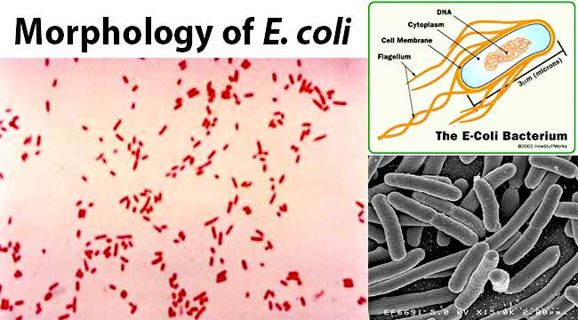


Escherichia Coli E Coli An Overview Microbe Notes


Academic Oup Com Labmed Article Pdf 32 7 368 Labmed32 0368 Pdf
1 Escherichia coli a Fix a smear of Escherichia coli to the slide as follows 1First place a small piece of tape at one end of the slide and label it with the name of the bacterium you will be placing on that slide 2Using the dropper bottle of deionized water found in your staining rack, place 1/2 of a normal sized drop of water on a clean slide by touching the dropper to the slideDifferential stains Gram stain In contrast to simple stains, differential stains are used to distinguish the difference between bacteria One of the most wellknown differential stains is Gram stain, which differentiates grampositive and gramnegative bacteria based on the difference in their cell wall structure The Gram stain was developed by the Danish bacteriologist Hans Christian GramE coli bacteria are a normal part of the intestinal flora in humans and other animals, where they aid digestion They are examples of bacilli shaped bacteria PASIEKA/Science Photo Library/Getty Images Bacillus Cell Arrangements Bacillus is one of the three primary shapes of bacteria Bacillus (bacilli plural) bacteria have rodshaped cells
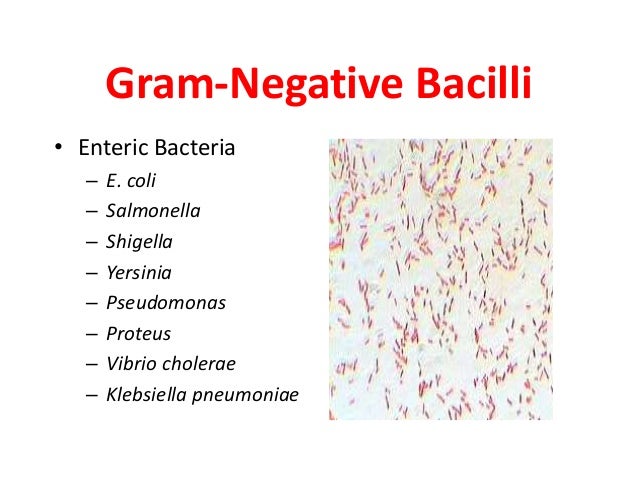


Basics In Identification Of Bacteria By Dr T V Rao Md


Gram Stain Of E Coli Bacterium A Gram Stain Of Shows Gramnegative Download Scientific Diagram
Escherichia coli colonies on eosinmethylene blue agar will have a characteristic green metallic sheen that other gramnegative bacteria will not have true when looking at a blood agar plate with bacterial growth, beta is complete lysis of the red blood cells, alpha is partial lysis, and gamma is no lysisWhat color is E coli when gram stained?Your unknown sample For example, when you perform a Gram stain, you will always include samples of Staphylococcus epidermidis (S epi), which is known to be Gram positive, and Escherichia coli, which is known to be Gramnegative If the Gram stain procedure works as it should, S epi will be purple and E coli will be pink


52 Microbiology Unknown Project Cscc Bio 2215 Ideas Microbiology Medical Laboratory Medical Laboratory Science


Laboratory Perspective Of Gram Staining And Its Significance In Investigations Of Infectious Diseases Thairu Y Nasir Ia Usman Y Sub Saharan Afr J Med
Jane Buckle PhD, RN, in Clinical Aromatherapy (Third Edition), 15 VancomycinResistant Escherichia coli Escherichia is a gramnegative bacterium, which under the microscope is shaped like a rod with a small tail It is widely distributed in nature (Brooker 08)Escherichia coli (E coli) is part of the normal intestinal flora Some strains are pathogenic and can cause gastroenteritis, UTIMany experiments in this course will utilize the Gram stain You should KNOW THE GRAM STAIN MATERIALS 1 Slant cultures of Escherichia coli, Micrococcus roseus, or other bacteria 2 Inoculating loops, Slides 3 Gram staining reagents in dropping bottles 4 Toothpicks 5 BHI plate from Experiment I PROCEDURE Heat Fixed Smear 1Simple stain streptobacillus, chains of rods Gram Stain (positive), purple
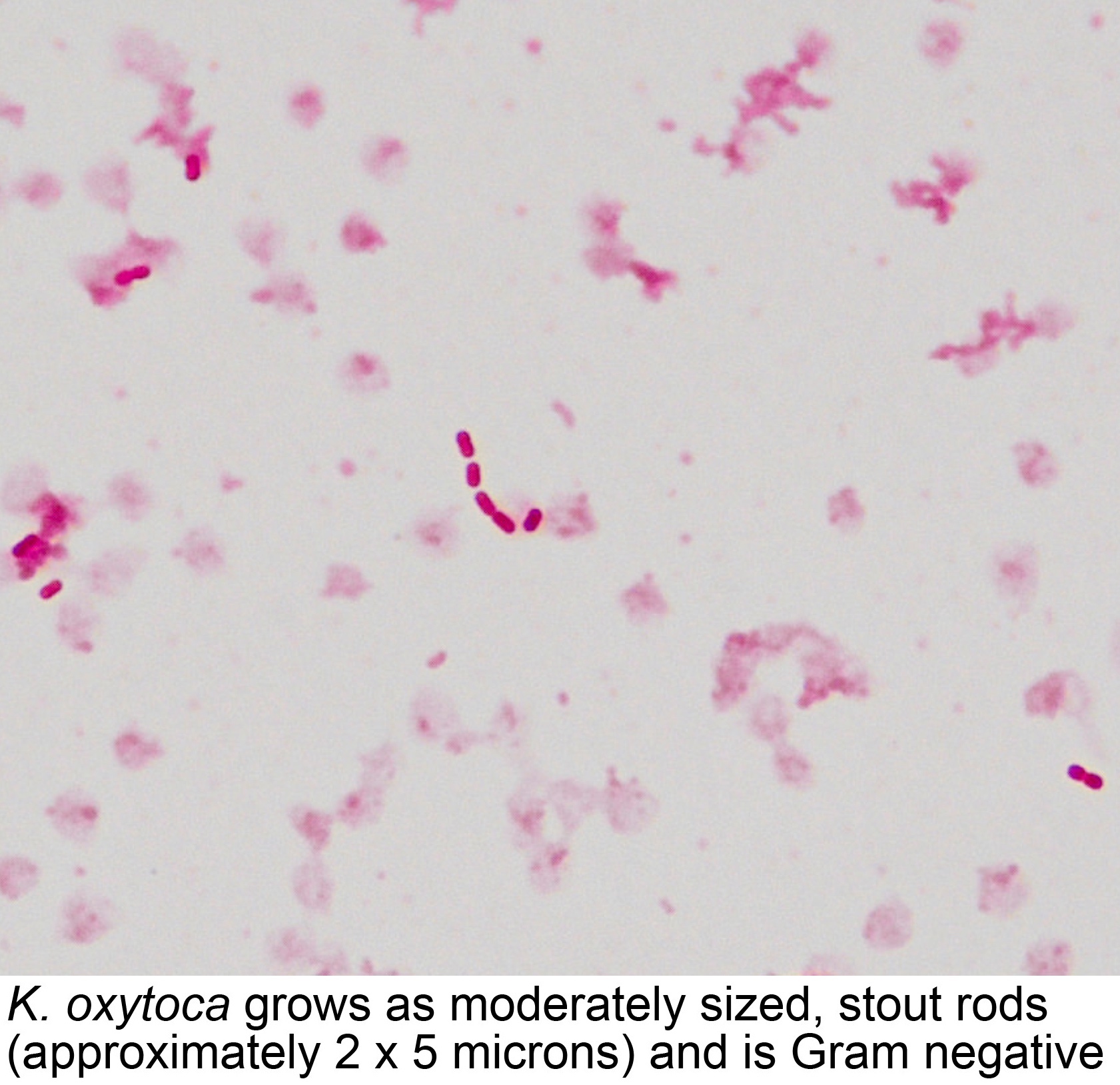


Pathology Outlines Klebsiella Oxytoca
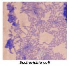


Microbiology Lab Exercise 6 Acid Fast Staining Flashcards Cram Com
Name the dye that gives it this color To what cell structure do the 2 dyes bind?Routine urine cultures should be plated using calibrated loops for the semiquantitative method Note The most commonly used criterion for defining significant bacteriuria is the presence of ⩾10 5 CFU per milliliter of urineWhen viewed under the microscope, Gramnegative E Coli will appear pink in color The absence of this (of purple color) is indicative of Grampositive bacteria and the absence of Gramnegative E Coli
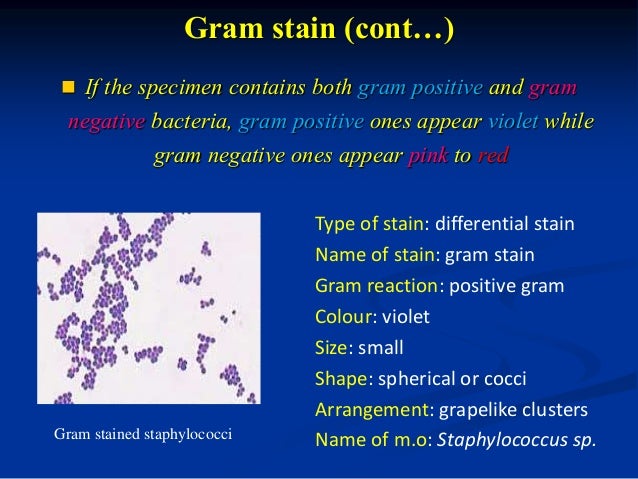


Bacterial Staining


Staphylococcus Aureus And Ecoli Under Microscope Microscopy Of Gram Positive Cocci And Gram Negative Bacilli Morphology And Microscopic Appearance Of Staphylococcus Aureus And E Coli S Aureus Gram Stain And Colony Morphology On Agar Clinical
Your unknown sample For example, when you perform a Gram stain, you will always include samples of Staphylococcus epidermidis (S epi), which is known to be Gram positive, and Escherichia coli, which is known to be Gramnegative If the Gram stain procedure works as it should, S epi will be purple and E coli will be pinkOn staining, E coli appear as nonsporeforming, Gramnegative rodshaped bacterium Routine urine cultures should be plated using calibrated loops for the semiquantitative method Note The most commonly used criterion for defining significant bacteriuria is the presence of ⩾10 5 CFU per milliliter of urine2 Morphology and Staining of Escherichia Coli E coli is Gramnegative straight rod, 13 µ x 0407 µ, arranged singly or in pairs (Fig 281) It is motile by peritrichous flagellae, though some strains are nonmotile Spores are not formed Capsules and fimbriae are found in some strains 3 Cultural Characteristics of Escherichia Coli


Gram Stain Of E Coli Bacterium A Gram Stain Of Shows Gramnegative Download Scientific Diagram


Gram Stain Lab Tests Online Au
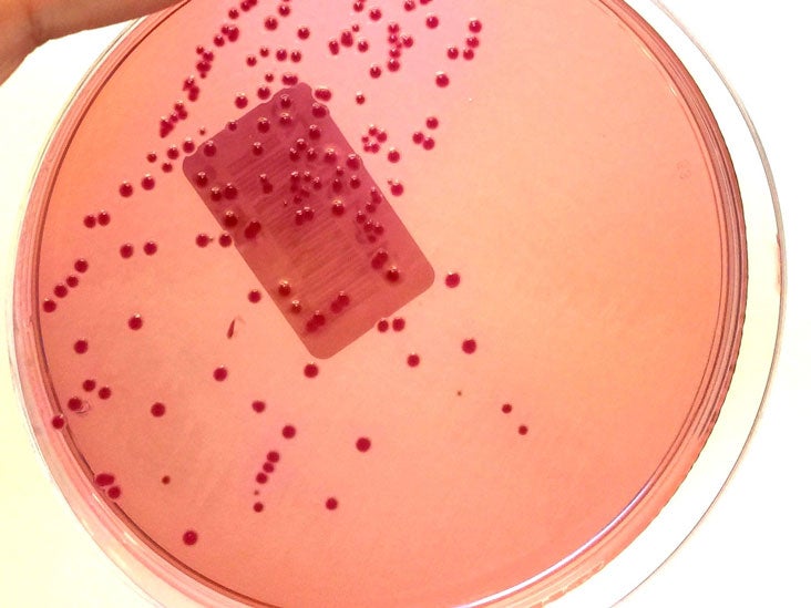
Cell structure, metabolism & life cycle E coli serotype O157H7 is a mesophilic, Gramnegative rodshaped (Bacilli) bacterium, which possesses adhesive fimbriae and a cell wall that consists of an outer membrane containing lipopolysaccharides, a periplasmic space with a peptidoglycan layer, and an inner, cytoplasmic membraneWhat color is E coli when gram stained?Escherichia coli Four different strains of Escherichia coli on Endo agar with biochemical slope Glucose fermentation with gas production, urea and H 2 S negative, lactose positive (with exception of strain D "late lactose fermenter";


What Does An E Coli Bacteria Look Like Under A Microscope Quora
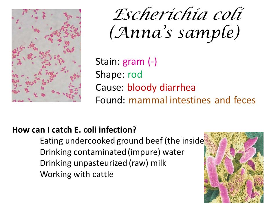


What Should You Have Seen Under The Microscope Activity Ppt Download
Mixed Ecoli & Saureus Gram stain, 1000x Mixed growth (cocci and bacilli) on BA from unwashed hand, Gram stain, 1000x Mixed growth (Gram pos & Gram neg) on BA from mouth swab, Gram stain, 1000xInclude shape and arrangement, as well as Gram stain result 6 Staining_Essay6_AcidFast E coli Image EXPERIMENT 2 Upload your acidfaststained EGram stain (negative), red Endospore stain vegetative cells, red What is the shape & arrangement of B cereus?
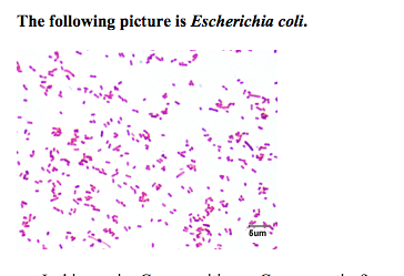


Solved Lab Module Staining Study Help Instructions Rea Chegg Com


Bacilli Wikipedia
Your unknown sample For example, when you perform a Gram stain, you will always include samples of Staphylococcus epidermidis (S epi), which is known to be Gram positive, and Escherichia coli, which is known to be Gramnegative If the Gram stain procedure works as it should, S epi will be purple and E coli will be pinkThese included smears made from pure broth cultures of Escherichia coli (E coli) and Staphylococcus aureus (S aureus) and a smear of a mixed broth culture containing E coli and S aureus Smears were heat fixed and gram staining conducted according to procedures outlined in the labster module on Gram Staning (Ref – Labster)What are the gram reaction, shape, and arrangement of E coli?


Microscopy And Staining


What Is The Escherichia Coli S Shape And Arrangement Quora
List at least 3 differences between gram positive and gram negative bacteria Would it be useful to perform a gram stain on a mixed culture?What are the stain results?What are the gram reaction, shape, and arrangement of E coli?


Morphology Culture Characteristics Of Escherichia Coli E Coli


Asmscience Examination Of Gram Stains Of Urine
On staining, E coli appear as nonsporeforming, Gramnegative rodshaped bacterium;Bio 112 Abstract Escherichia coli, Bacillus subtilis and Staphylococcus epidermidis were analyzed for this lab activity to determine their Gram Stain After the multilayered Gram Stain procedure each bacteria were classified as Grampositive or Gramnegative depending on their cell walls staining color The results showed that E coli stained pink and classified as GramnegativeCells are typically rodshaped, and are about μm long and 025–10 μm in diameter, with a cell volume of 06–07 μm 3 E coli stains Gramnegative because its cell wall is composed of a thin peptidoglycan layer and an outer membrane During the staining process, E coli picks up the color of the counterstain safranin and stains pink
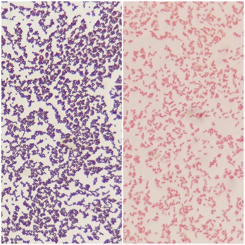


Pathogens Bacteria Viruses Gram Stain Teachmephysiology


Laboratory Perspective Of Gram Staining And Its Significance In Investigations Of Infectious Diseases Thairu Y Nasir Ia Usman Y Sub Saharan Afr J Med
Include shape and arrangement, as well as Gram stain result 5 Staining_Essay5_ E coli GramStain Classification EXPERIMENT 1 How would you classify the E coli bacteria after Gram staining?On Endo agar it looks like lactose negative)All four strains are mannitol positive (best seen in fig D), cellobiose negative (strains A, B)Cells are typically rodshaped, and are about μm long and 025–10 μm in diameter, with a cell volume of 06–07 μm 3 E coli stains Gramnegative because its cell wall is composed of a thin peptidoglycan layer and an outer membrane During the staining process, E coli picks up the color of the counterstain safranin and stains pink
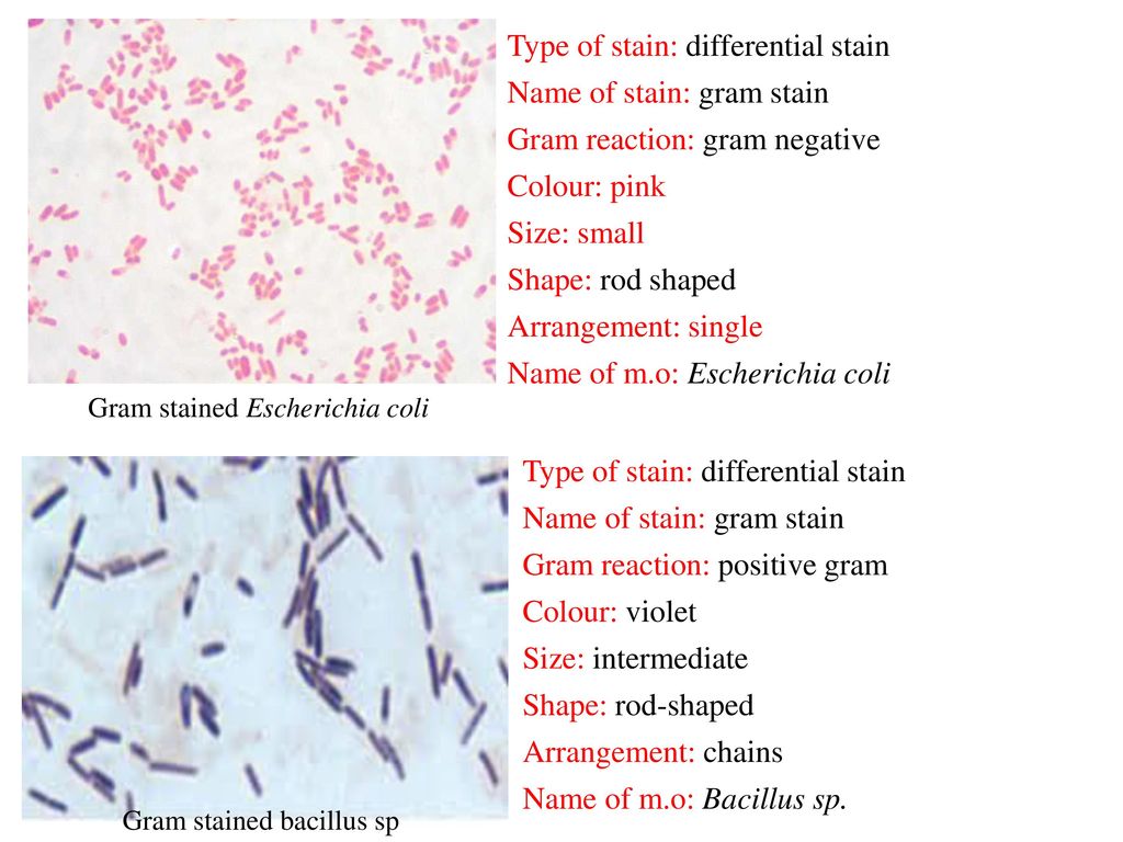


Gram Staining Principle Procedure And Results Msc Sarah Ahmed Ppt Download


Solved 1 Identify The Morphology Morphological Arrangem Chegg Com
Biology Of E Coli E coli ( Escherichia coli) are a small, Gramnegative species of bacteria Most strains of E coli are rodshaped and measure about μm long and 0210 μm in diameter They typically have a cell volume of 0607 μm, most of which is filled by the cytoplasmList at least 3 differences between gram positive and gram negative bacteria Would it be useful to perform a gram stain on a mixed culture?The colony morphology of organisms is observed Colony shape, size (in mm), color, consistency, elevation, opacity, and margin are observed and noted down for further identification Gram staining for identifying gram negative bacteria Gram staining is the beginning test in an identification procedure in bacterial classification


Bacteria Diversity Of Structure Of Bacteria Britannica


Q Tbn And9gcqykwkxckjvixk736ri4fxhsc0zoqnm21eamfac5sgematq0l I Usqp Cau
Classification on the basis of gram stain, bacterial cell wall, shape, mode of nutrition, temperature requirement, oxygen requirement, pH of growth, osmotic pressure requirement, number of flagella and spore formation (O1 is the pandemic strain) and E coli (enterotoxigenic, enteroinvasive, enterohemorrhagic,Escherichia coli Gram stained smear under microscopeRod shapedpink in colorthat's why Gram negative Bacilli#GramStain#GNB#GNRWhat are the gram reaction, shape, and arrangement of E coli?
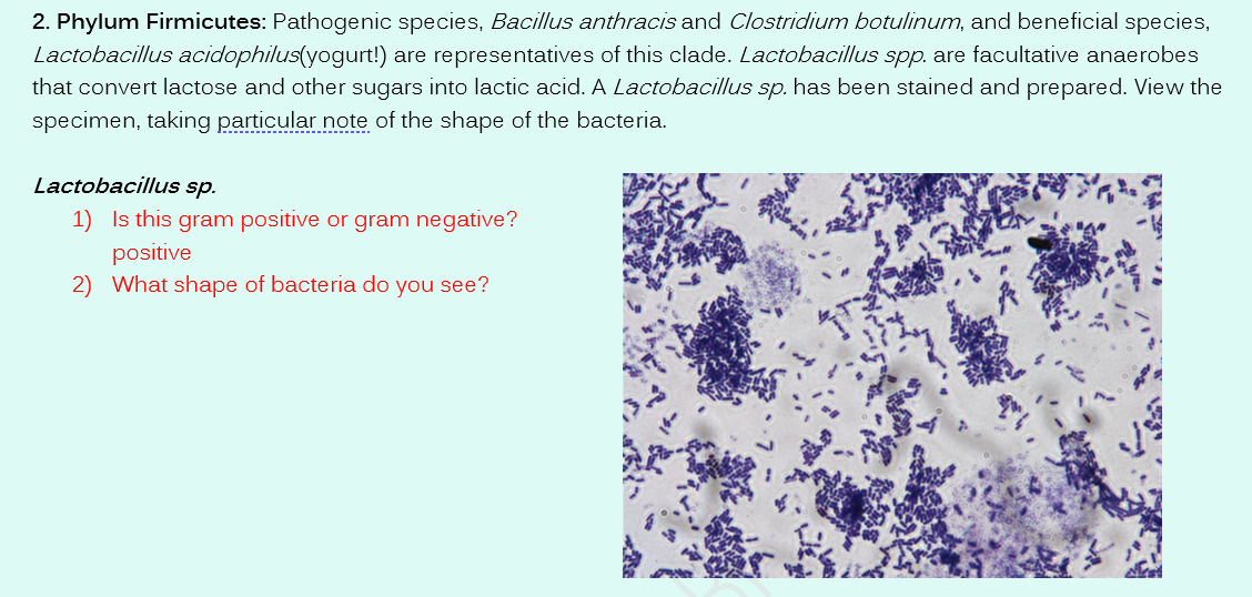


Solved 1 Phylum Proteobacteria The Largest Most Diverse Chegg Com


Identify The Stain And Shape Of The Bacteria Pictured Below Gram Positive Cocci Course Hero
The necks of the flasks Pasteur used were bent into an Sshape and left open to the air After Gram staining, E coli are pink in color This result is interpreted as _____ D a, c, b C All of the following are common to both the Gram stain and the acidfast stain EXCEPT A a decolorizing agent B a decolorizing agent and aDifferential stains Gram stain In contrast to simple stains, differential stains are used to distinguish the difference between bacteria One of the most wellknown differential stains is Gram stain, which differentiates grampositive and gramnegative bacteria based on the difference in their cell wall structure The Gram stain was developed by the Danish bacteriologist Hans Christian GramE coli is a facultative (aerobic and anaerobic growth) gramnegative, rod shaped bacteria that can be commonly found in animal feces, lower intestines of mammals, and even on the edge of hot springs They grow best at 37 C E coli is a Gramnegative organism that can not sporulate Therefore, it is easy to eradicate by simple boiling or basic


The Genesis Of Pathogenic E Coli Answers In Genesis
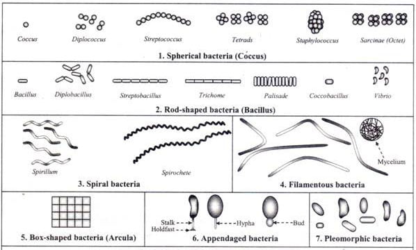


Different Size Shape And Arrangement Of Bacterial Cells
Colonies of some strains have typical greenish metallic sheen (figE) but many of them grow without it (figF) Escherichia coli (commonly abbreviated E coli) is a Gramnegative, rodshaped bacterium that is commonly found in the lower intestine of warmblooded organisms (endotherms)What are the stain results?Gram Staining Now that the slide has been heat fixed, you may now begin to stain the organism This is started by applying a few drops of crystal violet for 30 seconds, then rinsing with water for 5 seconds Next, cover the slide with Gram's Iodine and let sit for a full minute before rinsing with water for another 5 seconds
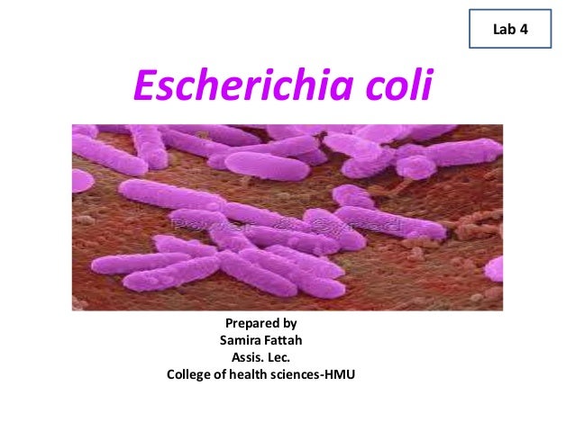


Escherichia Coli Lecture


Mixed Gram Positive And Gram Negative Bacillus W M Gram Stain Microscope Slide Science Lab Microbiology Supplies Amazon Com Industrial Scientific
Escherichia coli, often abbreviated E coli, are rodshaped bacteria that tend to occur individually and in large clumps E coli bacteria have a single cell arrangement, according to Schenectady County Community College E coli is a gramnegativE coli are classified as facultative anaerobes, which means that they grow best when oxygen is present but are able to switch to nonoxygendependent chemical processes in the absence of oxygen E coli bacteria are gramnegative, so they stain pink in a gram test E coli bacteria were first identified by bacteriologist Dr Escherich in 15Classification, Jejuni, Infection and Gram Stain Most of the bacteria that belong to this class are either spirilloids or curved in shape Family Like C coli, C jejuni is a foodborne pathogen which means that it is transmitted to an individual through contaminated food However, they can also be transmitted through contaminated
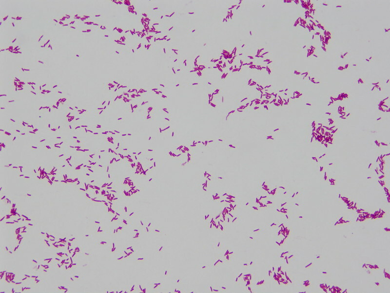


Pulsenotes Gram Negative Infections Notes


Optical Microscope Images Of E Coli Cells Following Gram Staining A Download Scientific Diagram
What color is E coli when gram stained?Gram positive bacteria stain bluepurple and Gram negative bacteria stain red The difference between the two groups is believed to be due to a much larger peptidoglycan (cell wall) in Gram positives


Identification Of S Aureus And E Coli From Clinical Specimens Download Table


Www Wiv Isp Be Qml Activities External Quality Rapports Atlas Bacteriology Gram Negative Aerobic And Facultative Rods Pdf
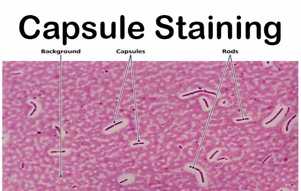


Capsule Staining Principle Reagents Procedure And Result


Gram Stains Of Bacteria Ppt Video Online Download


416pht Lab 2 Gram S Stain Acid Fast Stain Spore Stain Ppt Video Online Download


Photo Gallery Of Pathogenic Bacterial
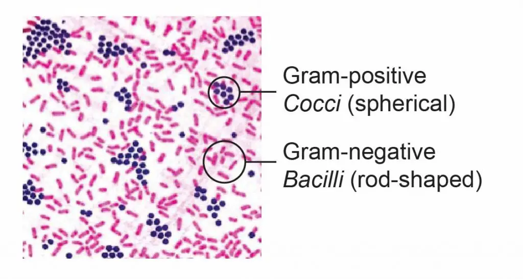


Observing Bacteria Under The Microscope Gram Stain Steps Rs Science


Bacteria 101 Cell Walls Gram Staining Common Pathogens Tusom Pharmwiki
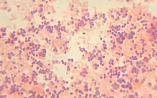


Staining Microscopic Specimens Microbiology


Www Wiv Isp Be Qml Activities External Quality Rapports Atlas Bacteriology Gram Negative Aerobic And Facultative Rods Pdf
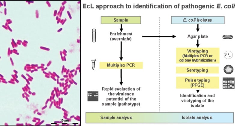


Escherichia Coli E Coli An Overview Microbe Notes


Escherichia Coli Wikipedia


Solved Lab Module 6 Gram Stain Lab Report Name Using T Chegg Com


Colony Characteristics Of Escherichia Coli Is A Gramnegative Facultatively Anaerobic Rodshaped Coliform Bacterium Of The Genus Escherichia That Is Commonly Found In The Lower Intestine Stock Photo Download Image Now Istock


Www Wiv Isp Be Qml Activities External Quality Rapports Atlas Bacteriology Gram Negative Aerobic And Facultative Rods Pdf


Academic Oup Com Labmed Article Pdf 32 7 368 Labmed32 0368 Pdf
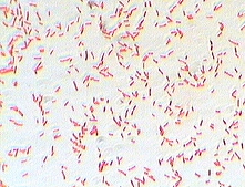


Photo Gallery Of Pathogenic Bacterial


Difference Between E Coli And Klebsiella Difference Between


Escherichia Coli E Coli Meaning Morphology And Characteristics


Chapter 2 Cells


Gram Negative Bacteria Shape Page 4 Line 17qq Com


Types Of Bacteria Information From Antibiotic Research Uk


E Coli Bacteria Britannica


What Does An E Coli Bacteria Look Like Under A Microscope Quora


Q Tbn And9gcru6r1unv3ndsfkjxwkvwwydqcu1p3g8xmz3ovuconfloxo8rwc Usqp Cau


Phenotypic Plasticity Of Escherichia Coli Upon Exposure To Physical Stress Induced By Zno Nanorods Scientific Reports


Laboratory Perspective Of Gram Staining And Its Significance In Investigations Of Infectious Diseases Thairu Y Nasir Ia Usman Y Sub Saharan Afr J Med


Www Mccc Edu Hilkerd Documents Bio1lab3 Exp 4 000 Pdf


The Lab Freaks Testing On Escherichia Coli


Laboratory Perspective Of Gram Staining And Its Significance In Investigations Of Infectious Diseases Thairu Y Nasir Ia Usman Y Sub Saharan Afr J Med


Gram Positive Cocci An Overview Sciencedirect Topics


Bacterial Characterization Using Gram Staining Figure 2 A The Download Scientific Diagram


Figure 3 From Pathologicalstudy On Colibacillosis In Chickens And Detection Of Escherichia Coli By Pcr Semantic Scholar
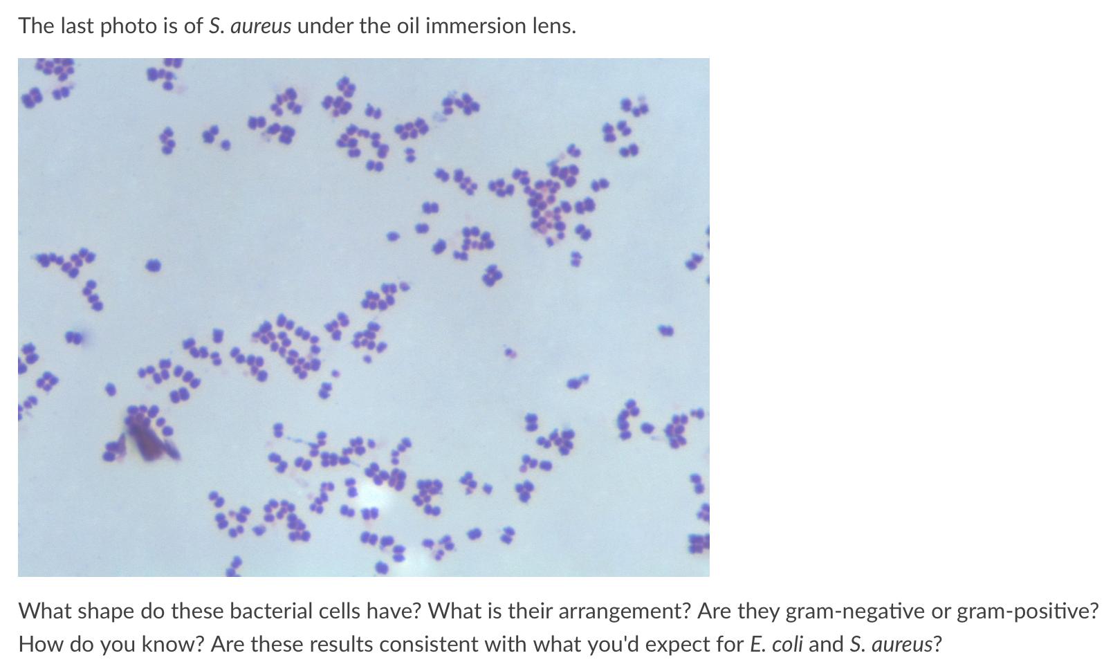


Solved The Last Photo Is Of S Aureus Under The Oil Immer Chegg Com


What Does Gram Positive Mean Fermup
/gram_positive_vs_negative-5b7f26d2c9e77c005746fbd7.jpg)


Gram Positive Vs Gram Negative Bacteria


Growth Requirements Of E Coli And Auxotrophs Microbiology Class Video Study Com
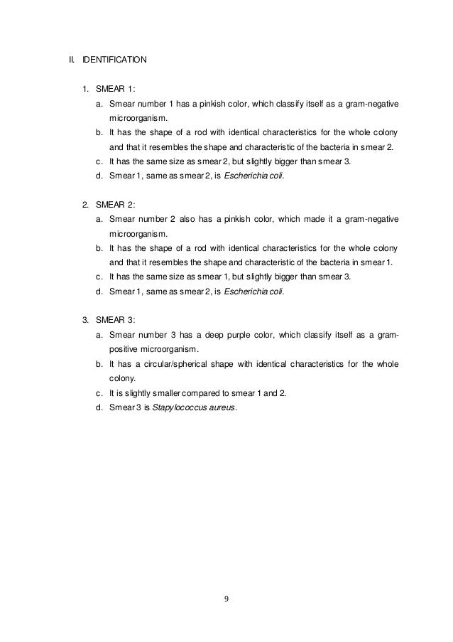


Lab Report Isolation Of Pure Culture Gram Staining And Microscopic


Gram Positive Bacteria Microbiology


Bacterial Staining


Module 7 8 10 Gram Stain Acid Fast Endospore Growth Characteristics Flashcards Quizlet


Www Wiv Isp Be Qml Activities External Quality Rapports Atlas Bacteriology Gram Negative Aerobic And Facultative Rods Pdf


Www Mccc Edu Hilkerd Documents Bio1lab3 Exp 4 000 Pdf


Gram Staining Procedure 42 Rights Managed Stock Photo Corbis Microbiology Medical Laboratory Educational Materials
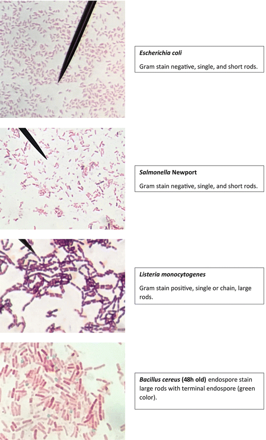


Staining Technology And Bright Field Microscope Use Springerlink


Path 417 Case 4 One Too Many Hamburgers Ppt Download
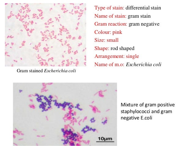


Bacterial Staining


Optical Microscope Images Of E Coli Cells Following Gram Staining A Download Scientific Diagram
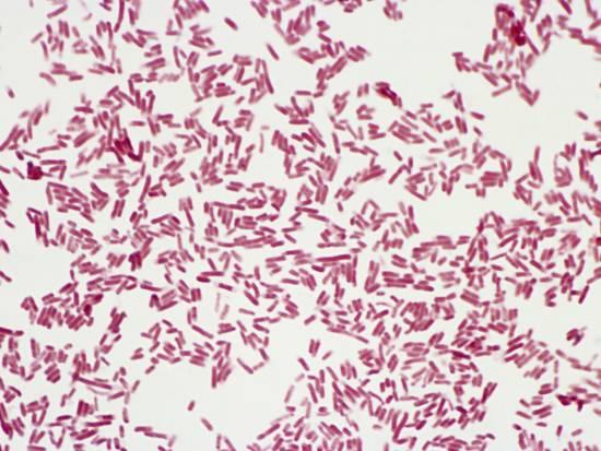


Biochemical Test Of Escherichia Coli E Coli Microbe Notes
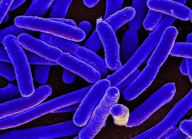


E Coli Under The Microscope Types Techniques Gram Stain Hanging Drop Method


Www Mccc Edu Hilkerd Documents Bio1lab3 Exp 4 Pdf


E Coli Gram Stain Introduction Principle Procedure And Result Interpret


Q Tbn And9gcqkye60ou Johpr02n Mbv1fferrjpdh Lnct7ymdf5qhyia1ld Usqp Cau


Photo Gallery Of Pathogenic Bacterial


Morphology Of Bacterial Cells
/gram-positive-staphylococcus-aureus-bacteria-541802136-57979cca5f9b58461f26eccc.jpg)


Gram Stain Procedure In Microbiology
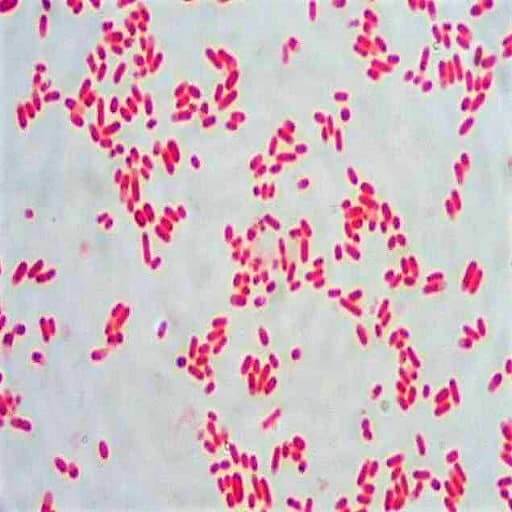


Morphology Culture Characteristics Of Escherichia Coli E Coli


Bacterial Characteristics Gram Staining Video Khan Academy


Gram Negative Bacteria Wikipedia


Bacterial Slides Bacerial Stains Microbiology


Escherichia Coli Colony Morphology And Microscopic Appearance Basic Characteristic And Tests For Identification Of E Coli Bacteria Images Of Escherichia Coli Antibiotic Treatment Of E Coli Infections


13 Macconkey Agar Ideas Microbiology Medical Laboratory Microbiology Lab
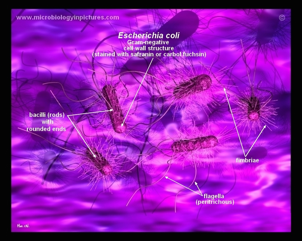


How E Coli Bacteria Look Like
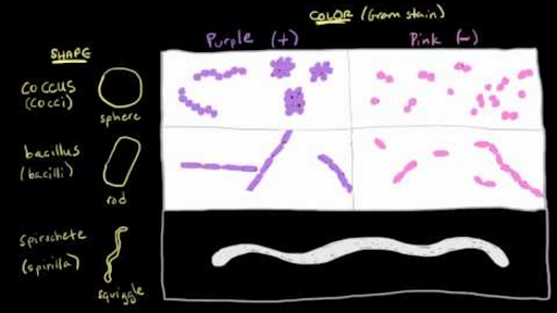


Bacterial Characteristics Gram Staining Video Khan Academy


Gram Stain Microbiology Lab


Gram Stain Wikipedia


Gram Negative Bacteria Images Photos Of Escherichia Coli Salmonella Enterobacter


Gram Stain Definition And Patient Education


Pin On Microbiology


Q Tbn And9gctnncfjtcedl5fo0rq5mdljtvlng Qopeaabn2fkjwz27muvuqg Usqp Cau


コメント
コメントを投稿